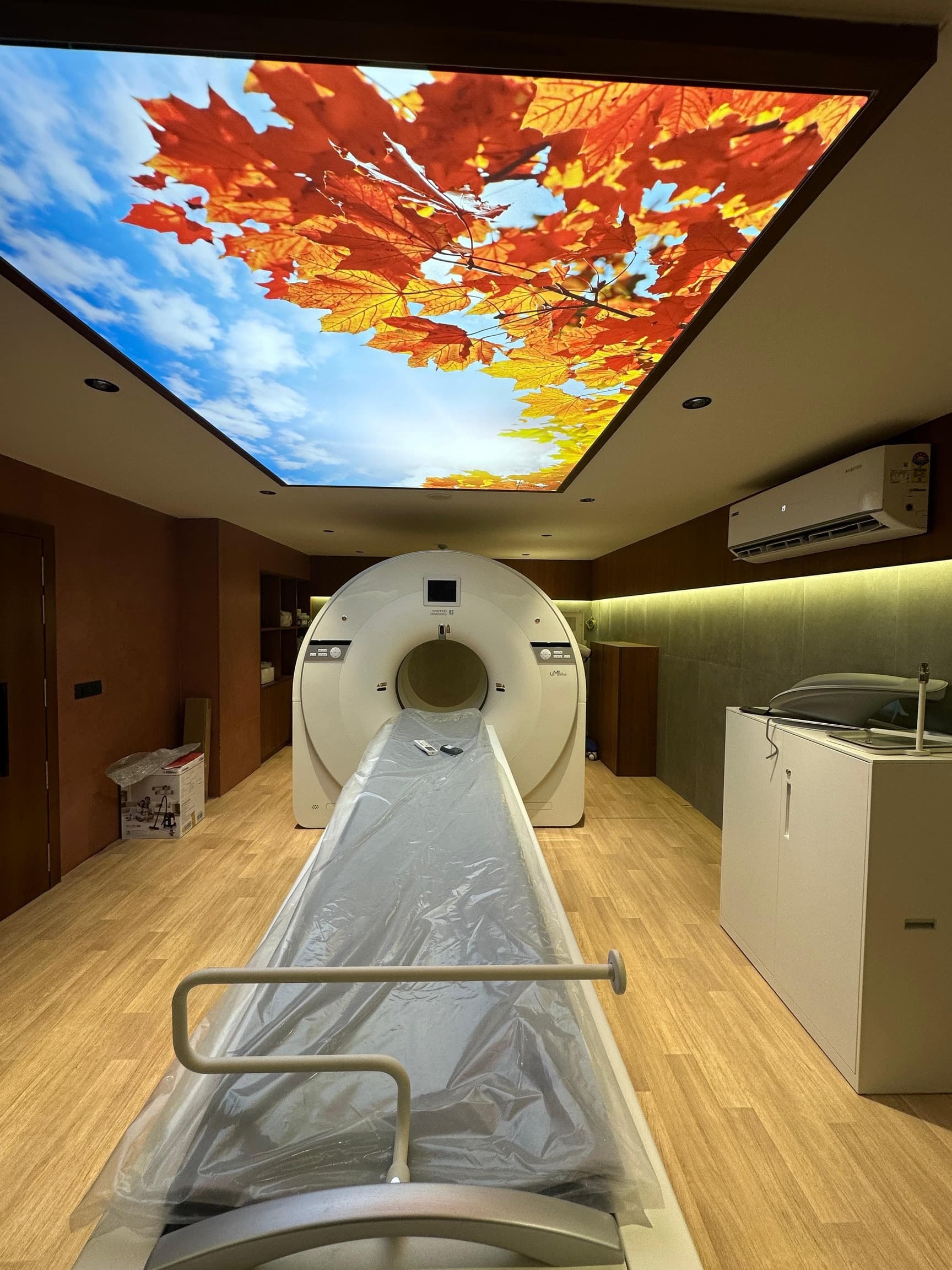
United Imaging uMI Vista
The only digital PET/CT in South Gujarat
The first and only digital PET/CT in South Gujarat — at the same price as a conventional scan.
The detector
Photon counting instead of guesswork
Most PET/CT scanners in India still use photomultiplier tubes — 1950s vacuum-tube technology that converts light into an electrical signal, losing a great deal of it along the way. The uMI Vista replaces those tubes with silicon photomultipliers: solid-state digital detectors that count individual light photons. More of the signal your body gives off actually reaches the image.
Every PET scanner does the same fundamental job: detect the faint flashes of light produced when the tracer in your body decays, and work out exactly where each flash came from. Resolution, radiation dose and scan time all follow from how well it does that.
With photomultiplier tubes, several crystals share one large detector and the position of each flash has to be estimated by comparing signals between neighbours. A great deal of information is lost in that estimation. With silicon photomultipliers, each crystal has its own detector and each photon is counted individually. Nothing is estimated.
What it delivers
Six things a digital detector changes
2.9 mm
spatial resolution
See smaller lesions
Roughly a third finer than a typical analogue scanner. Small nodes and early metastases that would otherwise blur into the background are resolved and reported.
~15 min
whole-body acquisition
Spend less time on the table
About half the usual scan time. Less movement blur, and a study that patients in pain or short of breath can actually complete.
Lower dose
protocols available
Take in less radiation
Higher sensitivity means we can achieve diagnostic images with a reduced injected dose — which compounds over a course of follow-up scans.
160
slice CT, 0.3 s rotation
Get a diagnostic CT too
The CT half is a full diagnostic scanner, not a low-dose localiser. One appointment, two complete studies.
Built in
cleaner images, faster
AI-assisted reconstruction
Deep-learning reconstruction suppresses noise without smoothing away the small lesions that matter, so lower-dose and shorter scans still read cleanly.
Same price
as a conventional PET/CT
Pay no more for it
We priced the advantage in, not on top. There is no reason to accept an older-generation scan to save money — because you would not be saving any.
Why 2.9 mm rather than 4–5 mm matters
Cancer staging is a set of thresholds, and thresholds are where resolution counts. Whether a single 6 mm lymph node is reported as involved can be the difference between a curative operation and starting with chemotherapy. Whether a solitary bone lesion is seen at all can decide between radical and palliative intent.
The scan is not just a picture. It is the evidence on which the treatment plan is built.
Side by side
Digital versus conventional PET/CT
The right-hand column is the typical range for the analogue, photomultiplier-based scanners still in routine service — a comparison of technology generations, not of any particular centre.
Detector technology
How the scanner turns your body's signal into a picture
Digital SiPM (silicon photomultiplier)
Analogue PMT (photomultiplier tube)
Digital detectors count light photon by photon instead of averaging them, so far less of the signal is lost before it becomes an image.
Spatial resolution
The smallest lesion the scanner can resolve clearly
≈ 2.9 mm
≈ 4 – 5 mm
Small deposits are found earlier — and in cancer, earlier detection is what changes the treatment plan.
Time-of-flight resolution
How precisely the scanner pinpoints where a signal came from
≈ 302 picoseconds
≈ 500 – 550 picoseconds
Sharper localisation means less blur and better contrast — the difference is most obvious in larger patients, where conventional images go soft.
System sensitivity
How much of the signal the scanner actually catches
≈ 12 cps/kBq
≈ 5 – 9 cps/kBq
Catching more signal is what allows either a shorter scan, a lower injected dose, or a sharper image — usually some of all three.
Scan time (whole body)
How long you lie still on the table
≈ 15 minutes
≈ 25 – 30 minutes
Shorter scans mean less movement blur — and are far kinder to patients in pain, breathless, or unable to lie flat for long.
Injected radiation dose
How much radioactive tracer has to go in
Reduced protocols routinely possible
Standard weight-based dose
A more sensitive detector needs less tracer for the same image quality — which matters most for young patients and anyone facing repeated follow-up scans.
CT component
The anatomical map PET is laid on top of
160-slice, 0.3 s rotation
16 – 64 slice, 0.5 – 0.8 s rotation
A faster, finer CT means crisper anatomy, fewer breathing artefacts, and a diagnostic-quality CT in the same sitting.
Bore & comfort
What it feels like to be scanned
Wide bore, short ring, ambient lighting
Narrower bore, longer acquisition
Nothing closes over your face. Claustrophobic and elderly patients get through the study far more comfortably.
uMI Vista figures are drawn from the manufacturer's published specification and from peer-reviewed NEMA NU 2-2018 performance assessment of the system. Individual results vary with patient size, protocol and clinical indication.
What this means for you
A clearer answer, the first time
Fewer equivocal reports means fewer repeat scans, fewer anxious weeks of waiting, and fewer unnecessary biopsies.
Kinder on the body
A shorter scan and a lower tracer dose matter enormously when you are already exhausted by treatment.
Treatment planned on better information
Correct staging is what decides between surgery, radiation and chemotherapy. A sharper scan makes that decision on firmer ground.
Same price. A generation ahead.
There is no premium for the digital scan. Call us and we will confirm the price for the study your doctor has requested.
3-4-5, Ground Floor, Zenon Building, Opp. Unique Hospital, Surat — Monday – Saturday, 8:00 AM – 8:00 PM