Whole-Body PET/CT (FDG)
18F-FDG
The complete head-to-thigh cancer survey — the single most useful scan in oncology.
≈ 15 min on the scannerView details
South Gujarat's first digital PET/CT
A PET/CT is a scan that shows how the cells inside your body are behaving — not just what they look like. Prismaa runs Surat's only high-resolution digital PET/CT — sharper images, half the scan time, and a lower radiation dose. At the same price as a conventional scan.
An X-ray or a CT scan is like a photograph of a city: you can see the buildings and the roads. A PET scan is like the same city photographed at night — now you can see which buildings still have their lights on. Cancer cells are the ones burning the most energy, so they light up brightest.
5,000+
Scans reported
Across PET/CT, CT and image-guided procedures
#1
In South Gujarat
The region's first and only digital PET/CT scanner
12
Specialists on the team
Radiologists, nuclear medicine physicians and operations
24 hrs
Typical report turnaround
Structured reports, most released within a day
4.9 from 120+ Google reviews — read what patients say
Understanding your scan
There are usually one of four reasons. Knowing which one applies to you makes the report much easier to understand when it arrives.
A lump on a CT scan may be a tumour, or scar tissue, or an old infection. PET shows whether it is metabolically active — and often spares people an unnecessary biopsy or operation.
Treatment for cancer confined to one organ is completely different from treatment for cancer that has travelled. One whole-body scan answers that in a single pass, instead of six separate tests.
A tumour can shrink and still be alive, or stay the same size and already be dead. PET shows which — usually months before size alone would tell you, so a failing regimen can be changed early.
After treatment ends, PET/CT can distinguish harmless scar tissue from disease that has come back, and can find recurrence while it is still small enough to treat aggressively.
What happens on the day
Around two and a half hours in the centre in total — most of it spent resting. The scan itself is the shortest part.
You come in having fasted for about six hours — water is fine and we want you to drink it. We check your blood sugar with a finger-prick, confirm your history and answer your questions.
Bring a relative. They can stay with you until the injection.
A tiny amount of a tracer — usually a sugar molecule carrying a safe, short-lived radioactive tag — goes into a vein in your arm. It is a single needle prick. You will not feel anything from the tracer itself: no warmth, no taste, no reaction.
The dose is small, and it clears from your body within hours.
You rest in a quiet room while the tracer travels through your bloodstream and collects in the cells that are working hardest. Staying still and relaxed matters here — moving muscles absorb tracer too, and that can blur the picture.
You can lie down. Most people doze off.
You lie on a padded table that moves slowly through a wide, short ring. It is open at both ends — nothing closes over your face. There is no noise beyond a soft hum, and no pain at all. On a conventional scanner this part takes 25–30 minutes; our digital scanner does it in about fifteen.
You can talk to the technologist at any moment. They can see and hear you throughout.
You eat normally straight afterwards and go home. We ask you to drink plenty of water to flush the tracer out, and to keep a little distance from pregnant women and young children for the rest of the day — a precaution, not a danger.
A fellowship-trained onco-radiologist reads your scan and issues a structured report written in the response criteria your oncologist uses. If you would like the findings explained face to face, we will sit down with you and your family and go through the images.
Ask us. Nobody should leave holding a report they cannot read.
The technology difference
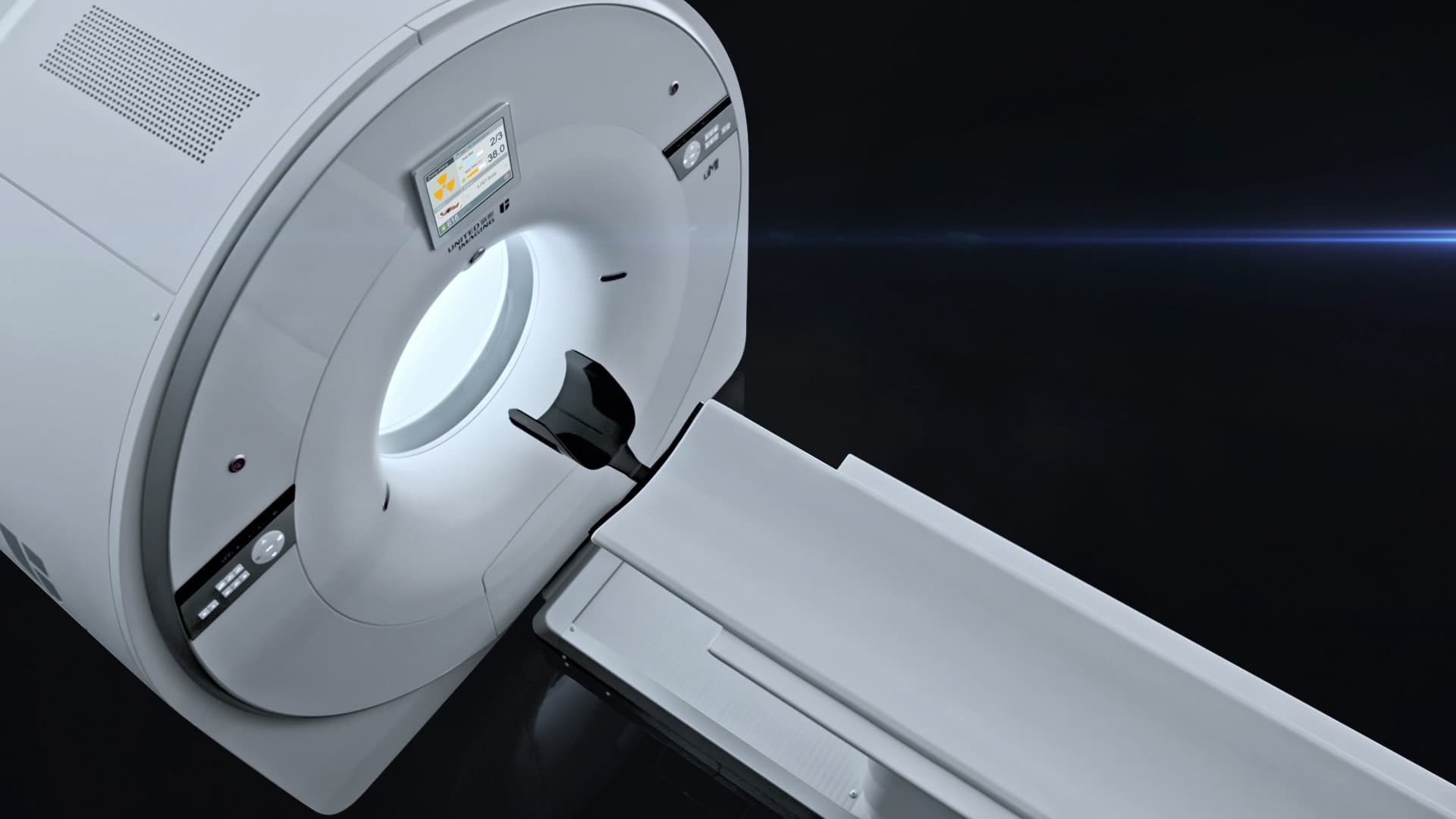
Most PET/CT scanners in India still use photomultiplier tubes — 1950s vacuum-tube technology that converts light into an electrical signal, losing a great deal of it along the way. The uMI Vista replaces those tubes with silicon photomultipliers: solid-state digital detectors that count individual light photons. More of the signal your body gives off actually reaches the image.
2.9 mm
spatial resolution
Roughly a third finer than a typical analogue scanner. Small nodes and early metastases that would otherwise blur into the background are resolved and reported.
~15 min
whole-body acquisition
About half the usual scan time. Less movement blur, and a study that patients in pain or short of breath can actually complete.
Lower dose
protocols available
Higher sensitivity means we can achieve diagnostic images with a reduced injected dose — which compounds over a course of follow-up scans.
160
slice CT, 0.3 s rotation
The CT half is a full diagnostic scanner, not a low-dose localiser. One appointment, two complete studies.
Built in
cleaner images, faster
Deep-learning reconstruction suppresses noise without smoothing away the small lesions that matter, so lower-dose and shorter scans still read cleanly.
Same price
as a conventional PET/CT
We priced the advantage in, not on top. There is no reason to accept an older-generation scan to save money — because you would not be saving any.
The right-hand column is the typical range for the analogue, photomultiplier-based PET/CT scanners still in routine service — a comparison of technology generations, not of any particular centre.
Detector technology
How the scanner turns your body's signal into a picture
Prismaa · Digital SiPM (silicon photomultiplier)
Conventional · Analogue PMT (photomultiplier tube)
Digital detectors count light photon by photon instead of averaging them, so far less of the signal is lost before it becomes an image.
Spatial resolution
The smallest lesion the scanner can resolve clearly
Prismaa · ≈ 2.9 mm
Conventional · ≈ 4 – 5 mm
Small deposits are found earlier — and in cancer, earlier detection is what changes the treatment plan.
Time-of-flight resolution
How precisely the scanner pinpoints where a signal came from
Prismaa · ≈ 302 picoseconds
Conventional · ≈ 500 – 550 picoseconds
Sharper localisation means less blur and better contrast — the difference is most obvious in larger patients, where conventional images go soft.
System sensitivity
How much of the signal the scanner actually catches
Prismaa · ≈ 12 cps/kBq
Conventional · ≈ 5 – 9 cps/kBq
Catching more signal is what allows either a shorter scan, a lower injected dose, or a sharper image — usually some of all three.
Scan time (whole body)
How long you lie still on the table
Prismaa · ≈ 15 minutes
Conventional · ≈ 25 – 30 minutes
Shorter scans mean less movement blur — and are far kinder to patients in pain, breathless, or unable to lie flat for long.
Injected radiation dose
How much radioactive tracer has to go in
Prismaa · Reduced protocols routinely possible
Conventional · Standard weight-based dose
A more sensitive detector needs less tracer for the same image quality — which matters most for young patients and anyone facing repeated follow-up scans.
CT component
The anatomical map PET is laid on top of
Prismaa · 160-slice, 0.3 s rotation
Conventional · 16 – 64 slice, 0.5 – 0.8 s rotation
A faster, finer CT means crisper anatomy, fewer breathing artefacts, and a diagnostic-quality CT in the same sitting.
Bore & comfort
What it feels like to be scanned
Prismaa · Wide bore, short ring, ambient lighting
Conventional · Narrower bore, longer acquisition
Nothing closes over your face. Claustrophobic and elderly patients get through the study far more comfortably.
Scans & services
Tap any scan to see what it is for, what it will tell your doctor, and how to prepare. PET/CT is not only for cancer — we also image the brain, the heart, and hidden infection.
18F-FDG
The complete head-to-thigh cancer survey — the single most useful scan in oncology.
68Ga-PSMA / 18F-PSMA
The gold standard for prostate cancer — finds disease that bone scans and CT miss.
68Ga-DOTA-TATE / TOC / NOC
For neuroendocrine tumours — pinpoints slow-growing tumours other scans overlook.
68Ga-FAPI
For breast cancer and solid tumours FDG struggles with — and it needs no fasting.
18F-DOPA
Maps the dopamine pathway — for brain tumours, movement disorders and neuroendocrine tumours.
68Ga-Exendin-4, 68Ga-Trivehexin, and others
Exendin-4 for insulinoma, Trivehexin and other targeted studies, arranged on request.
18F-FDG
Deauville-scored interim and end-of-treatment scans that guide chemotherapy decisions.
Flat-couch, laser-aligned scans that let the radiation oncologist target only what matters.
18F-FDG
Distinguishes Alzheimer's from other dementias by mapping how brain cells are working.
18F-FDG
Finds the seizure-generating zone in drug-resistant epilepsy, ahead of surgery.
18F-FDG
Answers the question before bypass surgery: is this heart muscle still alive?
18F-FDG
Weeks of fever with every test normal? PET/CT searches the whole body in one pass.
18F-FDG
Maps inflammation in blood vessels and organs, and tracks whether steroids are working.
Contrast, HRCT and ultra-low-dose protocols for chest, abdomen, brain and more.
Checks the heart's arteries for blockages — without a catheter in your groin or wrist.
CT-guided sampling of deep lesions — a definitive diagnosis without open surgery.
A specialist re-read of scans done elsewhere, with a structured, comparable report.
Advanced molecular imaging
Standard PET follows sugar, which every busy cell consumes. Gallium-68 tracers are built differently: each is designed to fit one protein found on one kind of tumour cell — so the cancer lights up and almost nothing else does.
Several cancers are poorly seen on a standard sugar-based scan — prostate cancer, neuroendocrine tumours, and pancreatic and peritoneal disease among them. A targeted tracer is often the difference between a scan that finds the disease and one that reports nothing abnormal.
These studies are produced to order, so please give us a few days' notice when booking.
68Ga-PSMA / 18F-PSMA
The gold standard for prostate cancer — finds disease that bone scans and CT miss.
Read more
68Ga-DOTA-TATE / TOC / NOC
For neuroendocrine tumours — pinpoints slow-growing tumours other scans overlook.
Read more
68Ga-FAPI
For breast cancer and solid tumours FDG struggles with — and it needs no fasting.
Read more
18F-DOPA
Maps the dopamine pathway — for brain tumours, movement disorders and neuroendocrine tumours.
Read more
68Ga-Exendin-4, 68Ga-Trivehexin, and others
Exendin-4 for insulinoma, Trivehexin and other targeted studies, arranged on request.
Read more
Beyond oncology
Roughly a fifth of the scans we perform are for something else entirely. If your neurologist, cardiologist or physician has asked for a PET/CT, this is why.
Neurology
MRI shows the brain's shape. PET shows how it is working — which is how Alzheimer's is told apart from frontotemporal or Lewy body dementia, and how the origin of drug-resistant seizures is located before surgery.
Learn moreCardiology
After a heart attack, weakened muscle may be dead scar or alive but starved. On an echo they look identical; they demand opposite decisions. PET tells them apart before bypass surgery is agreed.
Learn moreInfection & inflammation
White cells fighting infection burn sugar just as tumours do. One whole-body scan finds the hidden abscess, the inflamed artery or the occult lymphoma — and shows the physician exactly where to biopsy.
Learn more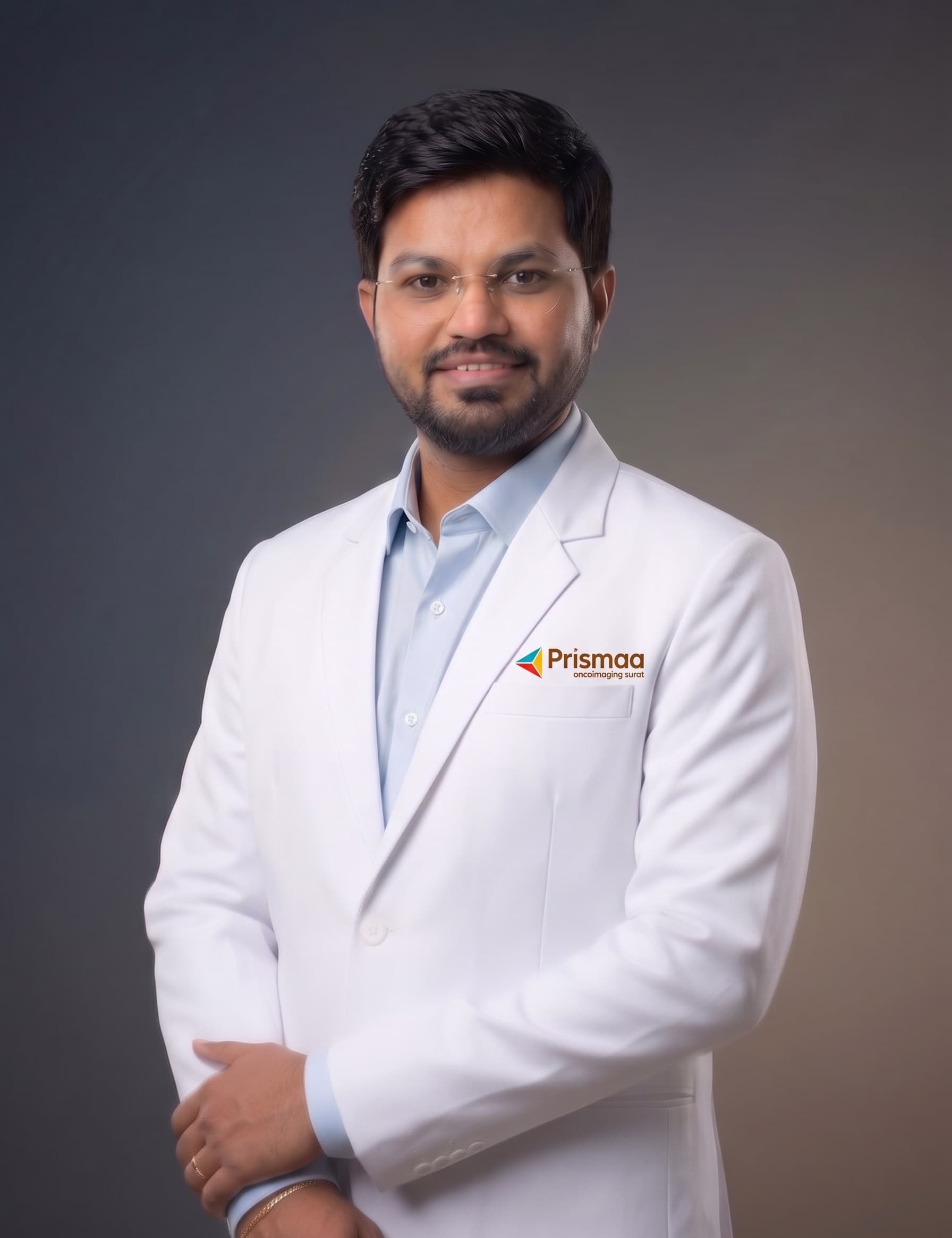
Who reads your scan
Founder, Managing Director & Lead Consultant Onco-Radiologist
MBBS, DNB (Radio-Diagnosis), Fellowship in Onco-Radiology
Dr. Keyur Mandaliya is an onco-radiology and molecular imaging specialist who founded Prismaa to bring current-generation digital PET/CT to Surat, so patients no longer need to travel to Ahmedabad or Mumbai for it.
He trained at Gujarat Imaging Centre (GIC PRIME), Ahmedabad, reporting on both conventional and digital PET/CT systems — experience that directly shaped Prismaa's decision to run a digital scanner. His practice covers cancer staging and response assessment (RECIST, PERCIST and related criteria), advanced tracer studies including PSMA, FAPI and DOTA-TATE, and image-guided biopsies, and he leads Prismaa's weekly tumour board.
Inside Prismaa
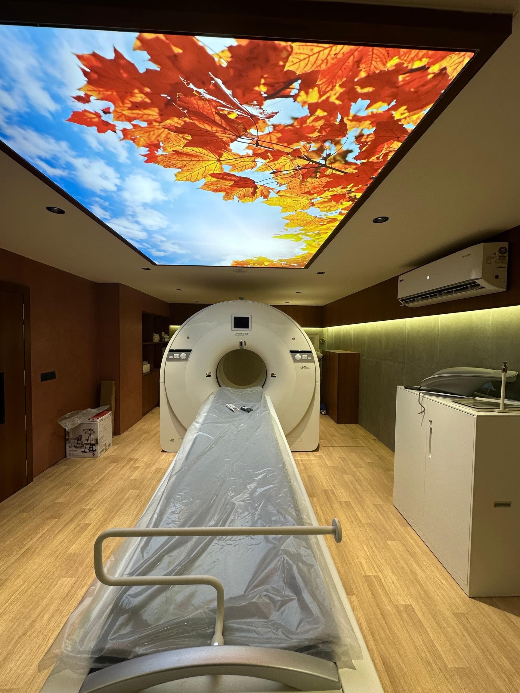
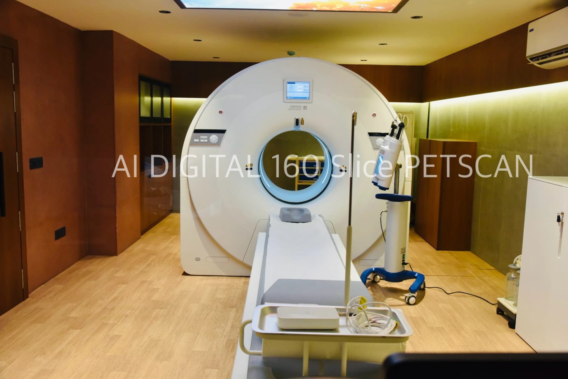
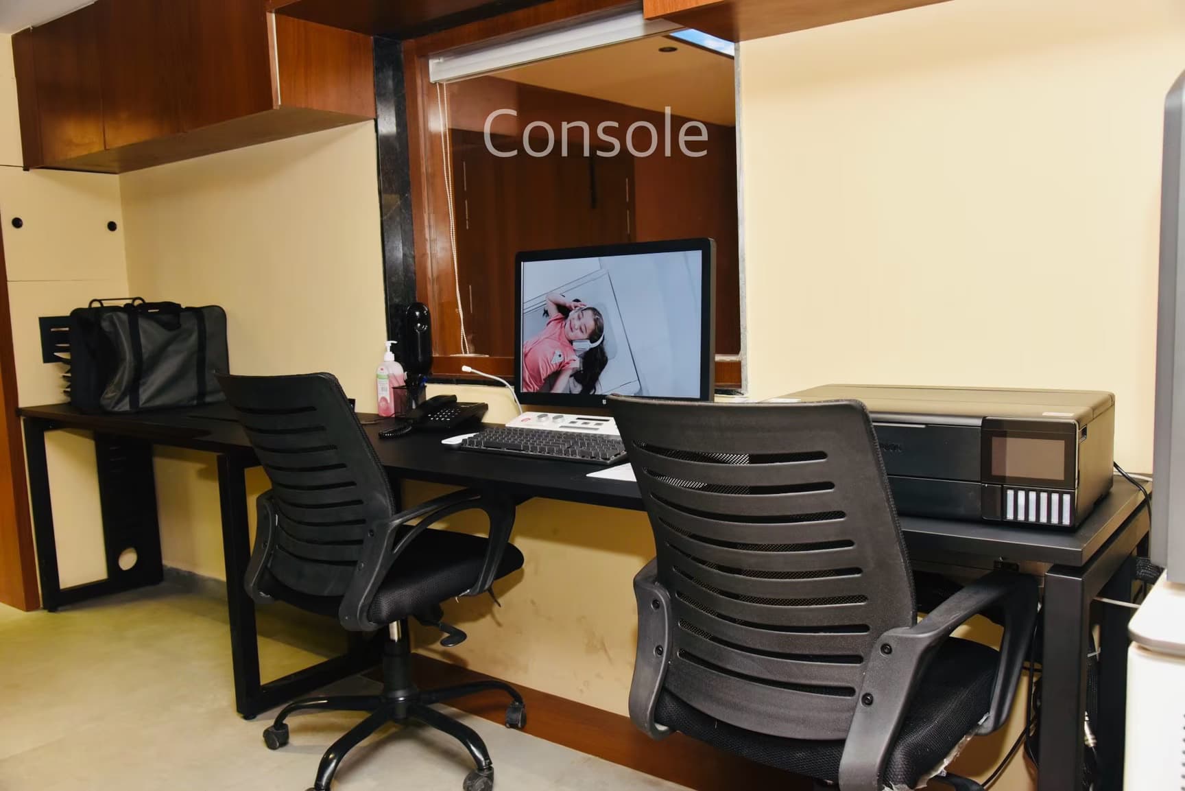
So we built somewhere that does not feel like a hospital corridor. Daylight, quiet private uptake rooms, and a scanner that is open at both ends.
In their words
The people who have been through it explain the experience better than we can.
We are filming patient stories now — in Gujarati, Hindi and English.
Until they are published here, the most honest picture of what it is like to be scanned at Prismaa is the unfiltered one: hundreds of reviews left by patients and their families on Google, which we do not curate or edit.
Patient guide
Plain-language explanations of PET scans, cancer, staging and treatment — written by our radiologists for patients and their families, not for other doctors.
Why PSMA PET has replaced bone scan and CT for prostate cancer staging, what a rising PSA after treatment means, and how it connects to Lu-177 therapy.
5 min read
How PET/CT is used in dementia, drug-resistant epilepsy, heart muscle viability and fevers of unknown origin — the non-oncological half of molecular imaging.
5 min read
Impression, SUVmax, FDG-avid, metabolically active, physiological uptake — the phrases that appear in every PET report, translated into English.
5 min read
TNM, stage groups and why staging decides your treatment. Written for patients and families who have just been given a number and no explanation.
5 min read
Silicon photomultipliers versus photomultiplier tubes, explained without the physics degree — and what the difference means for the person on the table.
4 min read
How much radiation a PET/CT actually delivers, how that compares to everyday life, who should avoid it, and how a digital scanner lowers the dose.
5 min read
Insights
Clinical notes, case observations and practice writing — some for patients, some for colleagues and researchers.
What we changed in reporting templates and workflow at Prismaa, what it did to turnaround time and report consistency, and what did not work.
4 min read4 August 2026
Lu-177 PSMA, PRRT and I-131 in plain language — how the same molecule can both find a cancer and deliver radiation to it, and who it is for.
5 min read14 July 2026
A practical note for referring clinicians on 68Ga-FAPI — the tumours where it outperforms FDG, why it needs no fasting, and how we are using it at Prismaa.
4 min read24 June 2026
Common questions
If your question is not here, call us. We would much rather explain it now than have you worry about it until the appointment.
Next at Prismaa
Imaging tells you where the disease is. The next step is treating it with the very same molecule — and we are building it.
We are commissioning a dual-head gamma camera and a dedicated radionuclide therapy suite. Together with our digital PET/CT, that will let one centre find the disease, confirm the target, and deliver the therapy — without patients travelling to another city between each step.
Bone scans, thyroid and renal studies, cardiac perfusion, sentinel node mapping and DMSA — the everyday nuclear medicine that Surat currently sends patients out of the city for.
Shielded therapy rooms for I-131 for thyroid cancer, Lu-177 PSMA for advanced prostate cancer, and Lu-177 DOTA-TATE (PRRT) for neuroendocrine tumours.
Scan with gallium-68, confirm the target lights up, then treat with lutetium-177 attached to the same molecule. See it, then treat exactly what you saw.
Tell us your diagnosis and we will let you know as soon as the relevant service opens.
Book a scan
Tell us the scan your doctor has asked for and we will confirm a slot, explain the preparation, and give you a clear price before you come in.